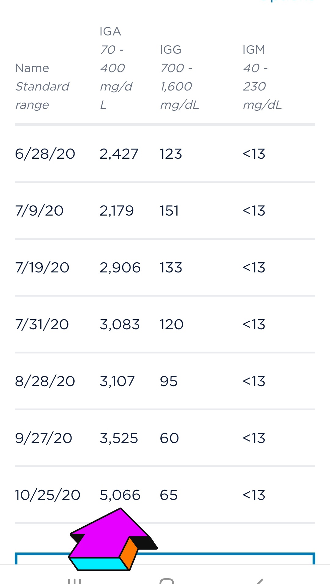
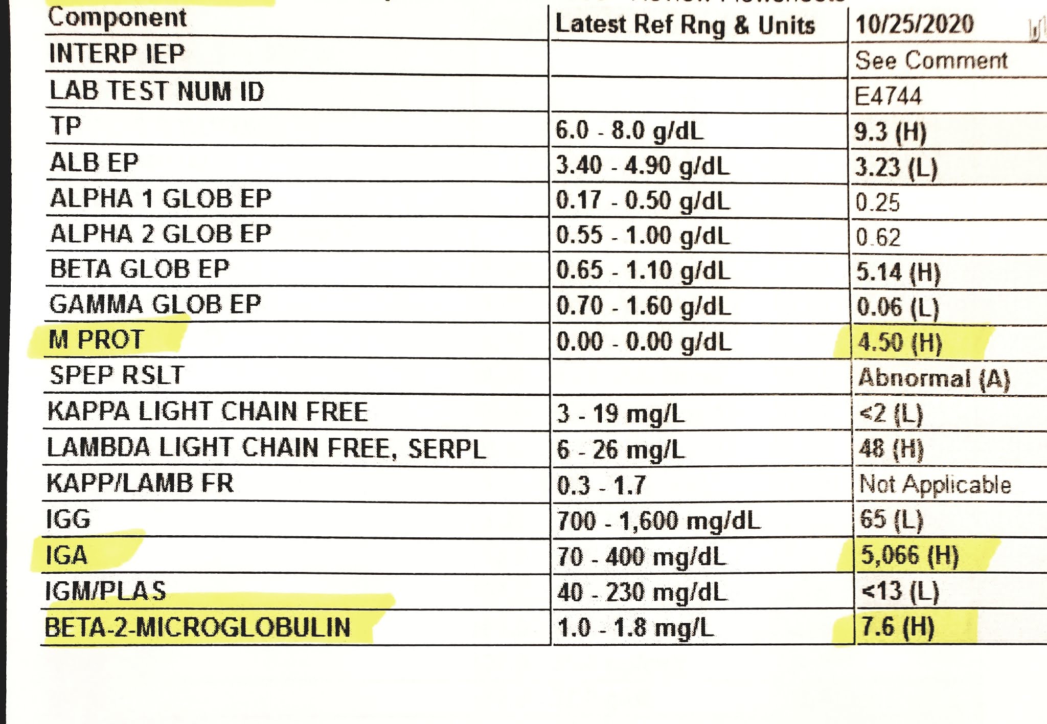
Cowgirl Up!!! ... Does Horse Poop Cause Cancer??

Friday, October 30, 2020

I'm So Sad... Scary Pet Scan Results

 10.30.20

Hello Friends-

I was just going to post my Pet Scan results from 10.24.20, (with little commentary), but then the irony of Halloween and Skeletons, bones, and spooky, scary results hit me. So here I am looking at the human skeleton and bones and organs and realizing how much I don't know about the human body. Well it seems that Myeloma wants to teach me a few lessons... Happy Halloween Julie, your skeleton is broken in so many areas. 


There are so many references to anatomy and physiology on my Pet scan and other reports, I have to always look up where things are. I didn't realize how far up our Rib Cage is. I didn't realize the placement of all of our internal organs. I always seem to think things are lower in our body then they actually are. Happy Halloween, my results are super spooky and scary :((

I've had Radiation all this week, except I cancelled on Wed, as I wasn't feeling good. Probably a combination of Mon and Tues chemo, steroid crash and (of course) new bone, muscle, nerve pain in my back and neck. I can never identify what type of pain it is. Can you? I never know if the pain and stiffness is from pulling, pinching something, or if I've cracked or fractured something again, or this time, if it's a side effect of the Radiation? This week is Radiation on my stinkn L side monster tumor that grew out of no where in summer. Remember the picture of that? Unbelievable. Well Radiation in July shrunk it a bit, but then it started to clunk into my L side neck, so my Radiation Oncologist wanted to radiate it again. My neck hurts, front and back, (muscles and nerves), and I've read I could have a sore throat for weeks, any time forward. Just never had so much pain in my life, and I can't stand it. I like being functional and happy, and pain steals that away :((


This monster is not that big on my chest anymore, but it's a big bump on the end of my L clavicle/collarbone. And so annoying when I move certain ways. It just grew so fast. I still blame Elotuzumab for all of this sudden insanity...

My L side middle-lower back hurts. Not sure if it's myeloma, or I pulled something, trying to do something around here. If I feel ok, (usually on steroid days), I want to be outside moving around, trying to do ranch chores etc, but I'm learning how limited I am, and trying to do anything I did before, has painful consequences. Just makes me so sad, as I used to think "retirement" would be so fun, just hanging out, taking care of our ranch, animals, plants, etc. That's all Jim and I wanted in our "Golden Years", along, of course, with being with family and friends.

Anyway, guess the Pet scan results were so alarming, my Oncologist called me yesterday morning and told me of all the new, or enlarged tumor, mass, fracture, etc areas. She spent a lot of time with me, explaining what the report said, and what my options could be. My Radiation Oncologist sent me a message that there are just too many areas to radiate, they can't radiate all of me... there's a progression of myeloma in multiple areas, etc. Bottom line, I have to brave up, and try new or more powerful chemos. Scary, so scary to me. Yes, Happy Spooky, Scary Halloween Julie. I hate, hate, hate new, unknown side effects. In the past, I always seemed to feel, do ok, on certain myeloma meds, even if my numbers were not good, but I did have an ok quality of life... I know too many myeloma patients that have done "aggressive treatments" (D-PACE?), and they are no longer here. Myeloma may appear to be "gone", then boom, they're "gone". I just want less pain, myeloma slowed down. I am not asking for an extreme result, and "cure" is not in my vocabulary at this time. I just want to live. I just want to feel ok. I'm not asking for my pre-myeloma life back. 

And here's my Scary IGA, M Protein, and Beta 2 results:
IGA Over 5000 now :((


Look at that crazy M Protein and Beta2!!! 


So to summarize, then I will post the entire report below... I have a new big mass near my R Kidney and Liver. It's big. They're worried about it. I'll have to have a biopsy on it. Ugh... ouch?? Makes sense tho, as I have had some dull and sharp pain on my R side for a while. It measures 6.1 x 9.4 x 9.2 cm!!! You can read the details in the report. My head is spinning... Seems as if all the masses, tumors, lesions, fractures, etc are larger, or growing, or more of them, etc. I just can't wrap my head around this. Will have to read and reread this. Amazing how smart and powerful Myeloma is, how it mutates, what it can do and does in our bodies. Why me tho. I am just so sad. I want to live...

Radiation, so amazing what is done...

Ok, here's the report. Thanks for reading and caring. Let me know your thoughts on all this. I just can't absorb it all. It can't be happening to me. I just keep thinking my body will "fix it" or it will turn out to not be happening to me. 


10/24/2020: PET SCAN: FINDINGS:

HEAD AND NECK:
Limited images of the brain demonstrate no focal asymmetric activity
to suggest metastatic disease. There is no midline shift or mass
effect within the calvarium. Please note PET/CT is suboptimal for
evaluation of intracranial metastatic lesions given intense
physiologic activity. Consider MRI of the brain to assess for brain
lesions, if clinically indicated. Last MRI brain 6/30/2020. Mild
heterogeneous activity of the clivus with limited evaluation due to
intense adjacent brain activity.

No hypermetabolic cervical lymphadenopathy is identified. Activity
of the vocal cords most likely phonation during FDG uptake. Bilateral
stable activity of the thyroid gland with SUV peak of 3 on the left
and 2.6 on the right.

CHEST:
The interval increase in size of the growth from the anterior aspect
of the right third rib expansile lesion now measuring 6.6 x 5.1 x 6.5
cm, compared to 3.5 x 2.8 cm on PET/CT from 5/7/2019. There is
circumferential increase in FDG activity with stable SUV peak of 4.4;
however, there is a low density center currently which is photopenic
on PET consistent with necrosis.

Interval increase in prominence of soft tissue nodule involving right
posterior third rib which now protrudes into the lung parenchyma
measuring 2.5 x 0.8 cm with SUV peak of 2.3 compared to 1.7
previously.

There is abnormal activity corresponding to pleural-based nodule
between the left 6th and 7th ribs posteriorly measuring 1.2 cm with
SUV peak of 2.8

Small bilateral pleural effusions. No hypermetabolic lymphadenopathy
in the mediastinum. No hypermetabolic lymphadenopathy in the
bilateral axilla or subpectoral regions. There is physiologic uptake
in the myocardium. No hypermetabolic focus bilateral breast
parenchyma.

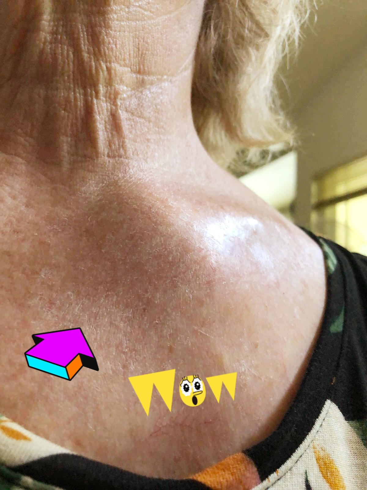
Intense abnormal activity of the left sternoclavicular joint region
with SUV peak of 4.5 compared to 2.9 previously.

Incidental finding of an azygos lobe, a normal variant.

ABDOMEN AND PELVIS:
Interval development of a large mass between the liver and the right
kidney measuring 6.1 x 9.4 x 9.2 cm with SUV peak of 4.5. There is
compression of the liver and the right kidney with this mass but
there appears to be at least a partial fat plane of separation
between the mass and the liver and the right kidney. The right
adrenal gland is not definitively visualized. There is photopenia of
the low-density lesion in the periphery of the right lobe of the
liver measuring 3 x 2.5 cm and left lobe of the liver measuring 1.3 x
1.6 cm. There is a round nonspecific focus anterior aspect of the
left lobe of the liver measuring 1.3 x 1.5 cm with SUV peak of 3.3
and hepatic lesion cannot be excluded. There is heterogeneous
activity in the remainder of the liver. There is a round focus of
intense abnormal activity in the liver, just anterior to the large
right upper quadrant mass, with SUV peak of 4.4..

There is mild activity in the stomach and heterogeneous activity
throughout the bowel. There are areas of segmental increase in FDG
activity throughout the bowel. Persistent activity in the anal region
with stable SUV peak of 4.3 compared to 4.2 previously.

Incidental note of cholelithiasis. Heterogeneous activity of the
spleen. No focal abnormal activity of the left adrenal gland. The
right adrenal gland is not definitively visualized. Low-density
lesion lateral aspect of the left mid abdomen measuring 2.2 x 1.6 cm
photopenic on PET.

There is physiologic excretion of FDG in the bilateral kidneys,
ureters and urinary bladder.

MUSCULOSKELETAL:
Evaluation of the osseous structures demonstrate interval increase in
abnormal activity of the lytic lesion at the right distal right
clavicle with SUV peak of 4.2 compared to 3 previously. Interval
increase in intense abnormal activity of the left sternoclavicular
joint region with SUV peak of 4.7 compared to 2.9 previously on
PET/CT from 5/7/2019. Interval increase in diffuse activity
throughout the spine. As a reference, L4 vertebral body activity has
SUV peak of 3.8 compared to 1.7 previously. L2 spinous process
abnormal activity has SUV peak of 3.8 compared to 2.5 previously.
Heterogeneous activity of the sternum with SUV peak of 3.1 compared
to 2.2 previously. Mild heterogeneous activity of the clivus.

UPPER EXTREMITIES:
Partial visualization of the upper extremities on CT demonstrate a
small focus of activity left distal humerus with SUV peak of 1.4.
Increase in activity bilateral wrists soft tissue distribution may
represent component of degenerative changes.

LOWER EXTREMITIES:
Interval increase in size and abnormal activity along the right
proximal femur with SUV peak of 4.1 compared to 2 previously.
Abnormal focus left proximal tibia with SUV peak of 3.4. Interval
increase in size, multiplicity and FDG activity of the right proximal
tibia shaft lucent lesion measuring 2 x 1.8 cm with SUV peak of 3.4
compared to 3.2 previously.


IMPRESSION:
Restaging PET/CT demonstrates unfortunate evidence for progression of
disease with interval increase in size of the growth from the
anterior aspect of the right third rib expansile lesion now measuring
6.6 x 5.1 x 6.5 cm, compared to 3.5 x 2.8 cm on PET/CT from 5/7/2019.
There is circumferential increase in FDG activity of the mass with
stable SUV peak of 4.4; however, there is a low density center
currently which is photopenic on PET consistent with necrosis.

Interval development of a large right upper quadrant mass measuring
6.1 x 9.4 x 9.2 cm with SUV peak of 4.5 located between the liver and
the superior aspect of the right kidney. The right adrenal gland is
not visualized.

Interval increase in prominence of soft tissue nodule involving right
posterior third rib which now protrudes into the lung parenchyma
measuring 2.5 x 0.8 cm with SUV peak of 2.3 compared to 1.7
previously.

Interval increase in abnormal activity of the lytic lesion at the
right distal right clavicle with SUV peak of 4.2 compared to 3
previously.

Interval increase in intense abnormal activity of the left
sternoclavicular joint region with SUV peak of 4.7 compared to 2.9
previously on PET/CT from 5/7/2019.

Several additional osseous findings: The spine, right femur,
bilateral tibia and sternum which have increased in FDG activity
compared to prior study, as detailed above. As a reference, diffuse
activity throughout the spine. As a reference, L4 vertebral body
activity has SUV peak of 3.8 compared to 1.7 previously.


Pet Scan 10.24.20

Stolen goals... 

Thank you for reading and caring as you do. 


4 comments:

  1. Hi Julie,
    After reading that PET report it is understandable why you are overwhelmed. Alot to absorb! You are brave Julie and you know you have to put your fear aside and get new stronger chemo in you!!! You can do it!! So the *&%$#* myeloma slows down at the least. Maybe bumping up the steroids too will help. Praying for you always. Sending you healing hugs. oxox

    ReplyDelete
    Replies
    1. Hi Bonnie, thank you so much for all your comments of support. Yes, the scan report is surreal, but I have to accept it's me, right. Thank you for thinking I am brave... I sure used to be, not so much any more. Just so tired of hurting and dealing with side effects. So may treatments I "could do", but not "brave enough to do so. Thank you friend, for all your support and correspondence xoxo

      Delete
  2. Hi Julie,
    A lot going on right now, isn't there? All we can really do is just put one foot in front of the other and keep going as best we can. Be gentle with yourself and lean on your supports when you need to. Hoping you find the right next step and sending my very best wishes for improvement in all test results, Bernadette

    ReplyDelete
    Replies
    1. Hi Bernadette, Thank you for your kind suggestions and support. Yes, one foot, one issue at a time. So hard for all of us that were always so busy and independent, but I have been humbled, and will move forward... slowly :))
      Hoping your status and treatment is going well friend, and you are feeling good. That's all I want for all my MM buddies. xoxo

      Delete

My Story... How my MM was diagnosed

October/November/December 2009...

Most of my life I was VERY presumptuous about being healthy, taking my (mostly) GOOD health for granted...
I was committed to annual check-ups for all of us, and so late October 2009, my daughter and I went for our annual and very routine physicals.

Surprise, surprise... my routine blood tests revealed extreme Anemia, significant White and Red Cell issues, low Platelets, and a variety of other CBC red flags! I was (stupidly) not worried when my GP doc left repeated phone messages to contact him, and when we did speak, I (stupidly) requested postponement of his referral appointment to the Hematology Dept until the end of the Fall academic term.

Arriving for my first appointment Dec 14, 2009, I was confronted with the check-in sign that read: "Hematology/Oncology"... What? Nooooo! not me... I must be in the WRONG place! And so my diagnosis journey began with vials and vials of blood drawn "stat", urgent Dr consultations, a surprise and painful Bone Marrow Biopsy, a full body Skeletal Scan, more blood tests stat, and then on 12.30.2009... THE revealing meeting... the "huh-what" moment ... the confirmation diagnosis that I, Julie, have CANCER!!!

Happy New Year to me, I just learned a new vocabulary word:
Multiple Myeloma!!! MM, Multiple Mye-what-loma!!!

January - June 2010

My medical metamorphosis began.
I read, and read, and read and researched and researched MM. I trusted my expert Oncology/Hematology team's plan and began my "New Normal" as a cancer patient.
My treatment plan was developed to include powerful Dexemthesone steroids paired with Revlimid chemotherapy, with the plan to be hospitalized for an Autologous Stem Cell Transplant July 2010.

I began living "one day at a time" like never before.
Jim was a wreck. Alissa and Scott were stunned; family and friends shocked.

Me... Cowgirl Up! I got back in the saddle and knew I was in for the ride of my life!
I did well on my initial pill-form Revlimid Chemo, "roid-rage" Dex Steroids and other supportive meds. I am forever deeply grateful and appreciative for all the love and support from everyone in my personal and professional life! I thank all of you for working along with me, and allowing me to continue to lead a semi "normal" life!
YOU have helped save my life!

My treatment trail ride forks to City of Hope hospital as I will saddle up beginning June 9, 2010 for a new rodeo called an Autologous Stem Cell Transplant!
Ye-Ha, let the adventure begin!

Chemical Warfare...

January 2010 - May 2010:
My initial chemo regimen:

Pill form Chemo= Revlimid (10mg, 15mg capsules)
Pill form Dexamethasone Steroids (40 mg, 4 days on, 4 days off!
Omeprazole for steroid acid reflux
Mepron (looks like yellow finger paint) Anti-fungal, Anti-viral, etc for my very compromised immune system
B-12
.81 Aspirin to prevent DVT, Revlimid complications
Allopurinol- keeping the kidneys healthy
Acyclovir- anti-Shingles, anti-viral

June 2010:
High dose IV Cytoxan chemo
Neupogen to build up stem cells for Apheresis, stem cell harvest, which was very successful, as City of Hope was able to collect 9.5 million of my own stem cells

July 2010 Hospitalization:
Two days of high dose Melphalan chemo
Then July 5, 2010 = my Autologous Stem Cell transplant infusion!

And you can read my whole story from that point forward in this blog!


What is multiple myeloma?

What is multiple myeloma?

Cancer starts when cells in the body begin to grow out of control. Cells in nearly any part of the body can become cancer, and can spread to other areas of the body. To learn more about how cancers start and spread, see What Is Cancer?

Multiple myeloma is a cancer formed by malignant plasma cells. Normal plasma cells are found in the bone marrow and are an important part of the immune system.

The immune system is made up of several types of cells that work together to fight infections and other diseases. Lymphocytes (lymph cells) are the main cell type of the immune system. The major types of lymphocytes are T cells and B cells.

When B cells respond to an infection, they mature and change into plasma cells. Plasma cells make the antibodies (also called immunoglobulins) that help the body attack and kill germs. Lymphocytes are in many areas of the body, such as lymph nodes, the bone marrow, the intestines, and the bloodstream. Plasma cells, however, are mainly found in the bone marrow. Bone marrow is the soft tissue inside some hollow bones. In addition to plasma cells, normal bone marrow has cells that make the different normal blood cells.

When plasma cells become cancerous and grow out of control, they can produce a tumor called a plasmacytoma. These tumors generally develop in a bone, but they are also rarely found in other tissues. If someone has only a single plasma cell tumor, the disease is called an isolated (or solitary) plasmacytoma. If someone has more than one plasmacytoma, they have multiple myeloma.

Multiple myeloma is characterized by several features, including:

Low blood counts

In multiple myeloma, the overgrowth of plasma cells in the bone marrow can crowd out normal blood-forming cells, leading to low blood counts. This can cause anemia – a shortage of red blood cells. People with anemia become pale, weak, and fatigued. Multiple myeloma can also cause the level of platelets in the blood to become low (called thrombocytopenia). This can lead to increased bleeding and bruising. Another condition that can develop is leukopenia – a shortage of normal white blood cells. This can lead to problems fighting infections.

Bone and calcium problems

Myeloma cells also interfere with cells that help keep the bones strong. Bones are constantly being remade to keep them strong. Two major kinds of bone cells normally work together to keep bones healthy and strong. The cells that lay down new bone are called osteoblasts. The cells that break down old bone are called osteoclasts. Myeloma cells make a substance that tells the osteoclasts to speed up dissolving the bone. Since the osteoblasts do not get a signal to put down new bone, old bone is broken down without new bone to replace it. This makes the bones weak and they break easily. Fractured bones are a major problem in people with myeloma. This increase in bone break-down can also raise calcium levels in the blood. (Problems caused by high calcium levels are discussed in the section “How is multiple myeloma diagnosed?”)

Infections

Abnormal plasma cells do not protect the body from infections. As mentioned before, normal plasma cells produce antibodies that attack germs. For example, if you developed pneumonia, normal plasma cells would produce antibodies aimed at the specific bacteria that were causing the illness. These antibodies help the body attack and kill the bacteria. In multiple myeloma, the myeloma cells crowd out the normal plasma cells, so that antibodies to fight the infection can’t be made. The antibody made by the myeloma cells does not help fight infections. That’s because the myeloma cells are just many copies of the same plasma cell – all making copies of the same exact (or monoclonal) antibody.

Kidney problems

The antibody made by myeloma cells can harm the kidneys. This can lead to kidney damage and even kidney failure.